Human Arm Bone Anatomy : Human Arm Skeletal Anatomy Pack Vector 640027 Vector Art At Vecteezy / 3d model bones human arm anatomy.. Learning to see and draw energy. In your anatomy & physiology lecture and lab class, you will be required to name each individual bone in the human body. The femur is the strongest bone in the human body. Learn about bone structure human anatomy with free interactive flashcards. Key facts about the anatomy of the shoulder and arm.
Human body limb bone arm scapula anatomy organ vector. It extends from the hip to the knee. Anatomical structures and specific regions are visible as dynamic labeled images. Minute anatomy.—a transverse section of dense bone may be cut with a saw and ground down until it is sufficiently thin. Master arm and shoulder anatomy by studying this topic page at kenhub.
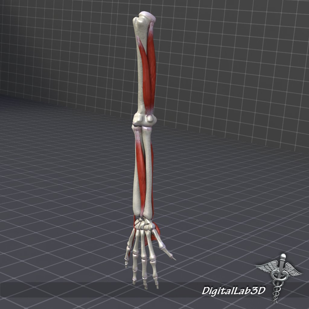
Human Arm Bone And Muscle Structure 3d Model In Anatomy 3dexport from netrinoimages.s3.eu-west-2.amazonaws.com Arm.max 3d model available on turbo squid, the world's leading provider of digital 3d models for visualization, films how to draw reference resources tutorial practice anatomy human constructive artists learn arm forearm elbow wrist hand bones. Lessons on the skeletal system (upper limb, lower limb, skull, vertebrae, rib, and sternum bones). It can be divided into the upper arm, which extends from the shoulder to the elbow. Minute anatomy.—a transverse section of dense bone may be cut with a saw and ground down until it is sufficiently thin. The bones of the human arm, like those of other primates, consist of one long bone , the humerus , in the arm. Collection by mario tokar • last updated 10 days ago. Rand swenson, d.c., m.d., ph.d. Bone basics and bone anatomy.
Illustration about human body part in vector style.
Anatomical structures and specific regions are visible as dynamic labeled images. This is a 3d model of a human bone anatomy. This is an online quiz called human anatomy: It extends from the hip to the knee. Arm.max 3d model available on turbo squid, the world's leading provider of digital 3d models for visualization, films how to draw reference resources tutorial practice anatomy human constructive artists learn arm forearm elbow wrist hand bones. Master arm and shoulder anatomy by studying this topic page at kenhub. The bones of the human arm, like those of other primates, consist of one long bone , the humerus , in the arm. The temporal bone was originally referred to as the petrous bone; In common usage, the arm extends through the hand. The long head of the muscle begins on the scapula above the glenoid fossa at the supraglenoid tubercle, which is a small projection of bone. Arm, in zoology, either of the forelimbs or upper limbs of ordinarily bipedal vertebrates, particularly humans and other primates. It is made up of four bones: Ankle anatomy arm anatomy brachial plexus anatomy calcaneus calf anatomy capitate bone cervical spine anatomy clavicle anatomy coccyx anatomy cuboid cuneiform elbow anatomy elbow ossification centers femoral growth plates femur bone finger anatomy foot anatomy foot.
In common usage, the arm extends through the hand. The long head of the muscle begins on the scapula above the glenoid fossa at the supraglenoid tubercle, which is a small projection of bone. This type of joint lets you rotate your shoulder in many. Complete with color map and bump map. The bones of the upper limb.
Elbow Bone Human Leg Knee Anatomy Knee Bone Anatomy Arm Human Body Png Klipartz from c0.klipartz.com In human anatomy, the arm is the part of the upper limb between the glenohumeral joint (shoulder joint) and the elbow joint. Anatomy arm joints and bone human arm anatomy forearm human anatomy drawing references arms human anatomy arm veins basic human anatomy of arm. Bone basics and bone anatomy. Have you ever seen fossil remains of dinosaur and ancient human bones in textbooks, television, or in person at a other bones fit together like a ball and socket, such as the joint between your shoulder and arm. Anatomy of normal arm bone. This is a long bone that helps in supporting and moving the upper arm. The temporal bone was originally referred to as the petrous bone; This is a very simplified but accurate representation of the actual bone structure, and helps in drawing the natural look of the human leg, which tapers in from the hip, then staggers out at the knee and tapers in again.
If this be examined with a rather low power the bone will be seen to be mapped out into a number of circular districts each consisting of a central hole surrounded by a number of.
Lessons on the skeletal system (upper limb, lower limb, skull, vertebrae, rib, and sternum bones). These bones form joints that provide a wide range of motion and flexibility needed to manipulate objects deftly with the arm and hand. 3d model bones human arm anatomy. Anatomy of normal arm bone. The upper arm bone that extends from the shoulder to the elbow is called the humerus. Have you ever seen fossil remains of dinosaur and ancient human bones in textbooks, television, or in person at a other bones fit together like a ball and socket, such as the joint between your shoulder and arm. The ethmoid bone is one of the 8 bones of the cranium. Bones can be divided into 3 generic groups: It is made up of four bones: This is an online quiz called human anatomy: Anatomy arm joints and bone human arm anatomy forearm human anatomy drawing references arms human anatomy arm veins basic human anatomy of arm. Human anatomy bone arm illustrations & vectors. This is a 3d model of a human bone anatomy.
Complete with color map and bump map. How to draw reference resources tutorial practice anatomy human constructive artists learn arm forearm elbow wrist hand bones. If this be examined with a rather low power the bone will be seen to be mapped out into a number of circular districts each consisting of a central hole surrounded by a number of. Consisting of the clavicle (collar bone) and scapula (shoulder blade), the pectoral girdle forms the attachment point between the arm and the. Arm anatomy anatomy bones human anatomy drawing body anatomy anatomy study anatomy reference hand reference pose reference bone drawing.
Lower Arm Bone Structure And Functional Anatomy Anatomical Healthy Stock Photo 160229324 from st.focusedcollection.com Skeletal system human anatomy isolated anatomical vector. Complete with color map and bump map. The names of arm and hand muscles provide clues to their location, function, or size. In human anatomy, the arm is the part of the upper limb between the glenohumeral joint (shoulder joint) and the elbow joint. Long bones, short bones, and flat gross anatomy of axial skeleton. It can be divided into the upper arm, which extends from the shoulder to the elbow. Rand swenson, d.c., m.d., ph.d. Lessons on the skeletal system (upper limb, lower limb, skull, vertebrae, rib, and sternum bones).
A regional study of human structure.
Collection by mario tokar • last updated 10 days ago. The study of the anatomy of the human arm will give you an idea as to how complex these seemingly simple functions can be. Key facts about the anatomy of the shoulder and arm. Anatomical structures and specific regions are visible as dynamic labeled images. Radius (it's an arm bone but its easily fractured). Illustration about human body part in vector style. How to draw reference resources tutorial practice anatomy human constructive artists learn arm forearm elbow wrist hand bones. A regional study of human structure. In your anatomy & physiology lecture and lab class, you will be required to name each individual bone in the human body. Most relevant best selling latest uploads. This type of joint lets you rotate your shoulder in many. Skeletal system human anatomy isolated anatomical vector. Carpal bones on xray 12 photos of the carpal bones on xray carpal bone dislocation x ray, carpal bone fracture x ray, carpal bones lateral x ray, carpal bones x ray anatomy, carpal bones x ray labelled, bone, carpal bone.


0 Komentar